RADIO VISION – SCIENTIFIC MILESTONE
Post Surgical Picture – Abdomen
The line of the incision is indicated by the Arrows A. B, C and X are blood vessels severed during the operation. The whitish effect below the line of the incision is due to inflammation. Arrow D shows a smaller blood vessel, also severed.
Hydatid Cyst in Liver
Cross section of patient’s liver, made through a blood crystal. The patient recorded a hydatid cyst or mole in diagnosis by Dr. Drown. The Radio-Vision Instrument was tuned in on the cyst, shown by Arrow A. The numerous other black circles and dots are bile ducts and blood vessels. The tissue structure should be compared with that shown in Plate 21, also a liver photograph.
Cancer Craters in the Liver
Arrows A and B show cancer craters in the liver of a patient residing in Indiana, the photograph being made in Hollywood, California from her blood crystal. The similarity of the liver tissue to that shown in Plate 20 should be noted. Arrow C shows a needle lesion in the patient’s liver from prior treatment.
Blood Clot and Cancer at Pyloric End of Stomach
This Radio-Vision photograph was made in England in 1939 by Dr. Ruth Drown, before an audience of seven British medical doctors, As she was discussing her work and methods, the patient’s blood crystal arrived from America, having originally been sent to her Holly wood office by Dr. Henry Pratt, a Connecticut osteopath. Symptoms included the patient’s inability to ingest or pass his food. Dr. Drown diagnosed the patient and stated that the man had cancer of the stomach and a blood clot recorded also. Tuning in on the blood clot, Dr. Drown made this picture in front of the M.D.’s. Arrow A shows the line of the pyloric end of the stomach, which extends from the right hand edge of the picture. Arrow B shows the white, thickened, cancerous tissue. Across the funnel-like opening of the stomach between A and B is the blood clot, C. The picture was sent out the same night on the trans—Atlantic clipper. Dr. Pratt received it shortly before the patient died. Post mortem surgery revealed the clot and the cancer to be exactly as shown in this photograph, taken from London, England, the previous night.
CONCLUSION
You have had your introduction to what is in all probability the greatest single invention of our time. You have seen with your own eyes photographs of the internal organs of the human body. You have seen photographs of various pathologies and foreign bodies. You have been presented with the evidence of the manner in which this instrument annihilates time and space.
The technology behind the Radio-Vision instrument exists as a successful system of diagnosis and therapy, it has been used and verified and welded into a useful system since Dr. Drown’s first invention in 1929. The Radio-Vision instrument has been in existence for twentyfive years, largely uncompre-hended in its significance and capacity to serve mankind.
To the reader who responds with “Has this been proven?” his attention is redirected to the photographs. Thereis the proof! There is no other way of making such photographs, and no way of “verifying” them, other than through the use of an inferior method.
Radio-Vision is a great diagnostic tool, a great investigative tool in other fields, a contribution to civilization and mankind’s march towards the light.
This booklet has been produced to acquaint physicians and intelligent laymen alike with the properties and performance of this Instrument. Many illusions exist concerning this work, which this book will help remove.
Radio-Vision is indeed a scientific milestone.

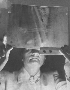
Dr. Ruth Drown studying a Radio-Vision photograph taken in New York of a patient in Wisconsin, using his blood crystallized as a tuner, diagnosis: abscessed tooth..
© Dr. Ruth B. Drown, 1960
- Dr. Ruth Drown studying a Radio-Vision photograph taken in New York of a patient in Wisconsin, using his blood crystallized as a tuner, diagnosis: abscessed tooth.


